Stone treatment can be divided into 3 main groups:
1. Medical treatment
2. Extracorporeal stone breaking (ESWL)
3. Surgical method (Endoscopic method – Open surgery method)
The main approach in stone treatment is; wait for small stones to pass by themselves, follow-up by doctor as long as they do not cause any harm (even if there is no pain, there may be permanent, even irreversible damage in kidneys), if the stone grows or if it was so big when it was first detected and it will not pass by itself, use extracorporal stone breaking method to break the stone. When there is a stone which is not suitable for breaking, endoscopic or open surgery removal of the stone should be tried.
A doctor should decide which stone treatment method will be preferred depending on the patient’s condition.
1. Medical TreatmentPractically is it is not possible to dissolve or break a kidney or urinary tract stone (excepet a very small group) by medicine. Medicine treatment can be used for; to help the passing of stones which are at the right size for passing (especially those in the ureter), prevent growing of small stones or prevent reformation of stone after the stones are treated in any method (in patients who carry the risk of stone reformation)
In stone passing treatment, the first stage is to decide which stone can pass and which method should be used for which stone by your doctor. Then, the treatment is proceeded according to stone location, the condition of your urinary tract.
In order to have medicine treatment for preventing growth of existing stones, first the reason for stone formation should be known. Some urine and blood tests can be required for this.
Protection treatment will be discussed in following sections.
2. Extracorporeal Stone Breaking Treatment: (ESWL – Extracorporeal Shock Wave Lithotripsy)
This is the most modern stone treatment method which is painless and which does not have the risk for surgery. It has been first clinically applied in 1980 in Germany.
It is based on the principle of focusing an extracorporally sourced high frequency sound waves (shock waves) on a stone and breaking the stone. The equipment may be different according to the shock wave producing method (piezo-electric, spark gap, electromagnetic) or stone imaging method (ultrasonography, x-ray).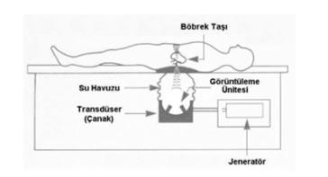
The shock waves focused on the stone cause surface erosion, and a stress wave which seperate structural elements of the stone and eventualy break the stone.
Previously, this method required a pool which the whole body is inserted in, ear protectors were used for protection from noise, and mostly anesthesia was necessary. Nowadays this method is very developed. The shock waves are transferred to body through a disk shaped tool or though a small pool where only the part of the body which the stone exist is inserted. There is no need for ear protectors. Anesthesia is very rarely necessary. Some advanced stone breaking equipments can also be safely used in baby-child patients. With the use of such equipments, the number of surgeries for stone treatment have been 90-95% decreased.
There may be differences in ESWL stone breaking equipments but they can be used for breaking stones anywhere in kidney, urinary tract and urinary bladder. For stones bigger than 3 cm, this method is not preferred as the first choice due to long treatment period, higher risk of complication. Complications (undesirable effects) of ESWL method are given below.
Hemorrhage area around the kidney (perirenal hematom):It is the most major complication. The occurrence percentage in small focused equipments is 0.6%, in big focused equipments 2-4%.
It is more common in patients with high blood pressure. Usually does not require any treatment, ESWL treatment should be suspended until the hemorrhage area is completeley healed. Rarely, the hemorrhage is not confined to kidney area and continues to enlarge. In these cases open surgery may be required.
Exposure to radiation:Not appliacble in equipments which has ultrasonographic imaging.
Bruise and tissue hardening (fibrosis) in kidney and in surrounding area due to shock wave. More common in big focused equipments, frequent and long treatment periods.
Stone pieces blocking the urinary tract (Ureter obstruction): The broken stone pieces may cause blockage in urinary tract. Sometimes a big stone at the front and sand following it may deposit. (Stone road) This case which is more common with big stones may be treated by breaking the big stone piece at the front.
3. Surgical Method
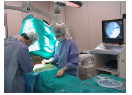
Surgical method can be simply divided into two groups; open surgery and removing the stone by endoscopic method.
The endoscopical method is becoming more important with advances especially in endoscopic equipments and increase in surgical experiences. Removing the stone by endoscopical method is done by either going in through the body’s natural openings (through the last prar of the urinary tract) or making a 1cm long cut in the skin and reaching the stone by going through the tissues (PCN- perkütan nefrolitotripsi). In order to remove the stone (big stone), once the stone is reached it may have to broken down. In this appliaction, depending on the size and location of the stone different stone breaking methods (mechanical, electrohydraulic, laser) can be used. There may be a possibility to continue with open surgery in each endoscopic operation.
 In open surgery method, a surgical operation is done at an area depending on the location of the stone. It is the most accurate method in stone treatment. It has become less common due to risk of anesthesia, cut pain, risk of infection, constriction in the urinary tract, risk of urine leakage, long term confinement to bed. Nowadays, it is preferred in treatment of very big stones which are not suitable for other treatments, and cases which there are other structural anomalies in addition to stone.